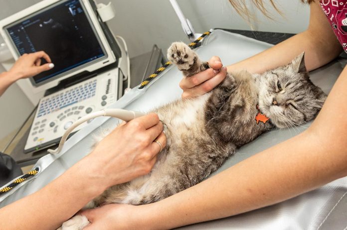
An ultrasound examination (AKA echography, ultrasonography) is a non-invasive imaging technique that allows internal body structures to be seen by recording echoes or reflections of ultrasonic waves. Unlike x-rays, which are potentially dangerous, ultrasound waves are safe. Ultrasound is considered the best, non-invasive and non-painful way to evaluate fluid-filled and soft tissue organs in pets.
These organs include the pet’s liver, spleen, kidney, pancreas, eyes, lymph nodes, testicles, intestinal tract, prostate, uterus/ovaries and heart. In more advanced equipment it is possible to evaluate the movement of the heart, the blood flow, the speed of the blood and calculate blood pressure.
Ultrasound equipment directs a narrow beam of high-frequency sound waves into the area of interest. The sound waves may be transmitted through, reflected or absorbed by the tissues that they encounter. Then the ultrasound waves that are reflected will return as “ultrasound echoes” received by the probe, and then converted into images through a sophisticated computer program. Ultrasound is also fast. Depending on the area, scanning usually takes approximately 5-10 minutes at most, and results may be seen immediately on a monitor and captured digitally.
The technique is excellent for examination of internal organs in the abdomen and chest. Ultrasound can diagnose problems earlier, finding internal abnormalities: the presence of nodules, masses, free fluid, cysts and abscesses, so it can lead to more successful treatment outcomes.
Ultrasound is painless and non-invasive, using sound waves that are virtually risk-free. Ultrasound can detect a problem deep within tissue or an organ without invasive surgery. If a sample of tissue is needed, an ultrasound-guided biopsy can be performed after your pet has been given appropriate sedatives and/or anesthetics.
Anesthesia is rarely needed because the technique is painless and most of pets feels comfortable in the dark room where the scan is being performed. Occasionally, if the patient is very frightened or fractious, a sedative may be necessary.
In some cases, with long hair dogs or cats, the fur must be clipped to perform an ultrasound exam and allow a perfect contact between the probe and the skin of the pet.
And while the initial cost of a scan may seem high, the value of the information obtained towards a final diagnosis in many cases saves time and resources, and overall, improving the chances for the right treatment for the pet.


